41 brain diagram and labels
Brain: Ultimate Guide to the Brain for AP® Psychology - Albert The forebrain consists of the thalamus, hypothalamus, amygdala, and the hippocampus. The hypothalamus, amygdala, and hippocampus make up what we call the Limbic System of your brain. Thalamus The thalamus is located between the cerebral cortex and the midbrain. It is made up of nuclei that receive different sensory and motor inputs. Parts of the brain: Learn with diagrams and quizzes | Kenhub Labeled brain diagram First up, have a look at the labeled brain structures on the image below. Try to memorize the name and location of each structure, then proceed to test yourself with the blank brain diagram provided below. Labeled diagram showing the main parts of the brain Blank brain diagram (free download!)
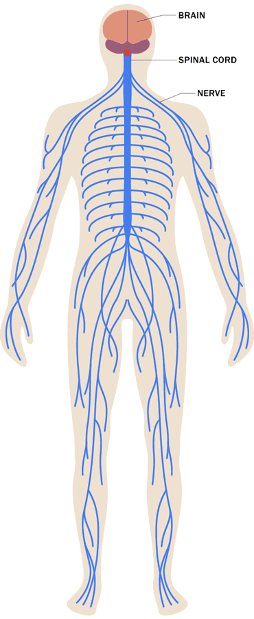
Human brain - Wikipedia The human brain is the central organ of the human nervous system, and with the spinal cord makes up the central nervous system.The brain consists of the cerebrum, the brainstem and the cerebellum.It controls most of the activities of the body, processing, integrating, and coordinating the information it receives from the sense organs, and making decisions as to the instructions sent to the ...

Brain diagram and labels
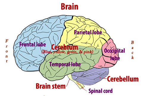
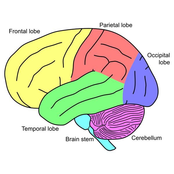
Parts of the Human Brain | Anatomy & Function - Study.com Each hemisphere of the outer layer of the cerebrum, the cortex, is composed of four lobes: the frontal lobe, the parietal lobe, the temporal lobe, and the occipital lobe. The four lobes of the... Brain Anatomy: Lesson for Kids - Video & Lesson Transcript | Study.com This part of the brain is responsible for muscle control, balance, and coordination. It helps you with everything from playing sports to writing your homework. Diagram showing some of the main ... Ventricles of the Brain Function, Anatomy & Diagram - Study.com A schematic diagram of the brain, with the lateral ventricles labeled. The Four Brain Ventricles The four main brain ventricles include the two lateral ventricles, the cerebral aqueduct, and third ...
Brain diagram and labels. Cross-sectional anatomy of the brain - e-Anatomy - IMAIOS Anatomical parts Angular gyrus Anterior cerebral artery Anterior commissure Anterior limb of internale capsule Anterior lobe of cerebellum Anterior quadrangular lobule [H IV and H V] Aqueduct of midbrain; Cerebral aqueduct Archicortex Atrium - Lateral ventricle Basilar artery Basilar plexus Biventral lobule ( H VIII) Body of caudate nucleus Positions and Functions of the Four Brain Lobes - MD-Health.com The brain is divided into four sections, known as lobes (as shown in the image). The frontal lobe, occipital lobe, parietal lobe, and temporal lobe have different locations and functions that support the responses and actions of the human body. Let's start by identifying where each lobe is positioned in the brain. Position of the Lobes › en › e-AnatomyAnatomical diagrams of the brain - e-Anatomy - IMAIOS These original illustrations and diagrams of the brain were created from 3D medical imaging reconstructions and then redrawn and colored using Adobe Illustrator. ... use of interactive anatomical labels. The user can select to display multiple categories of labels on the illustrations: Cerebral lobes / regions; Cerebrum, divided into: frontal ... Anatomy of the Brain - Simply Psychology The four lobes of the brain are the frontal, parietal, temporal, and occipital lobes (Figure 3). Figure 3. The cerebrum is divided into four lobes: frontal, parietal, occipital and temporal. Frontal lobes The frontal lobes are located at the front of the brain, behind the forehead (Figure 4).
Central nervous system: Anatomy, structure, function | Kenhub They are enveloped and protected by three layers of meninges, and encased within two bony structures; the skull and vertebral column, respectively. The brain consists of the cerebrum, subcortical structures, brainstem and cerebellum. The spinal cord continues inferiorly from the brainstem and extends through the vertebral canal. Brain: Function and Anatomy, Conditions, and Health Tips Some of its main functions include: processing sensory information regulating blood pressure and breathing releasing hormones Brain diagram Use this interactive 3-D diagram to explore the brain.... developingchild.harvard.edu › resources › the-brainThe Brain Circuits Underlying Motivation: An Interactive Graphic The brain systems that govern motivation are built over time, starting in the earliest years of development. These intricate neural circuits and structures are shaped by interactions between the experiences we have and the genes we are born with, which together influence both how our motivation systems develop and how they function later in life. Lobes of the brain: Structure and function | Kenhub The lobes of the cerebrum are actually divisions of the cerebral cortex based on the locations of the major gyri and sulci. The cerebral cortex is divided into six lobes: the frontal, temporal, parietal, occipital , insular and limbic lobes. Each lobe of the cerebrum exhibits characteristic surface features that each have their own functions.
Human Brain Lesson for Kids: Function & Diagram - Study.com It even helps you understand what's going on around you by receiving messages from your senses: touch, taste, smell, sight, and hearing. The Cerebellum Another part of your brain is called the... byjus.com › biology › liver-diagramLiver Diagram with Detailed Illustrations and Clear Labels Liver Diagram The liver is one of the most important organs in the human body. Anatomically, the liver is a meaty organ that consists of two large sections called the right and the left lobe. Brain MRI: How to read MRI brain scan | Kenhub In case you have identified any alteration in the signals coming from the brain tissue, it's useful to determine its location with respect to the lobes of the brain, since some pathological conditions tend to happen in specific lobes. There are six lobes of the brain; frontal, temporal, limbic, parietal, insular and occipital lobe s. Illustrations and diagrams of the 12 pairs of cranial nerves - IMAIOS This human anatomy module is about the cranial nerves. It consists of 15 vector anatomical drawings with 280 anatomical structures labeled. It is intended for the use of medical students working on human anatomy, student nurses, physiotherapists, electro-radiological technicians and residents - especially those working in neurology, neurosurgery, otolaryngology - and for any physician ...
32 Blank Brain Diagram To Label - Labels Database 2020
What are the 12 cranial nerves? Functions and diagram The cranial nerves are a set of twelve nerves that originate in the brain. Each has a different function responsible for sense or movement. The functions of the cranial nerves are sensory, motor ...
Brain Diagram - Cliparts.co
byjus.com › biology › diagram-of-heartHeart Diagram with Labels and Detailed Explanation - BYJUS The diagram of heart is beneficial for Class 10 and 12 and is frequently asked in the examinations. A detailed explanation of the heart along with a well-labelled diagram is given for reference. Well-Labelled Diagram of Heart. The heart is made up of four chambers: The upper two chambers of the heart are called auricles.
Human Anatomy for health & wellness Unit Plan
14 Informative Facts, Diagram & Parts Of Human Brain For Kids Human brain and the spinal cord are covered by three layers of tissue collectively known as the meninges. A clear fluid called cerebrospinal fluid flows between these layers (10) (11). The left part of the brain is responsible for analytical thinking. The right part helps in creative thinking (12).
Brain Viewed from Above | ClipArt ETC
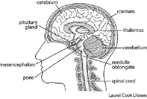
wikieducator.org › Nervous_System_Worksheet_AnswersNervous System Worksheet Answers - WikiEducator Jan 14, 2008 · 8. The diagram below shows a section of a dog’s brain. Add the labels in the list below and, if you like, colour in the diagram as suggested. Cerebellum - blue; Spinal cord - green; Medulla oblongata - orange; Hypothalamus - purple; Pituitary gland - red; Cerebral hemispheres – yellow. 9. Match the descriptions below with the terms in the list.
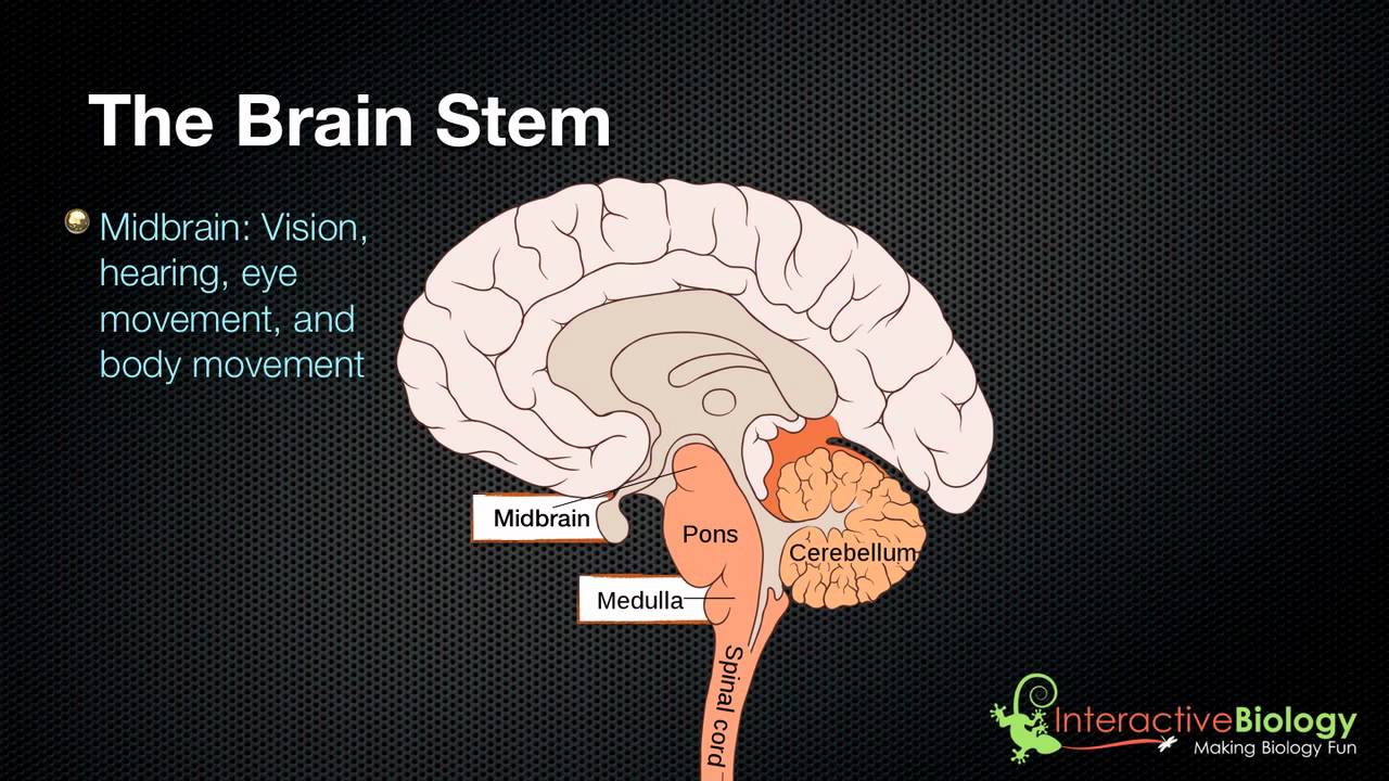
027 The 3 parts of the brain stem and their functions - YouTube
Labeled imaging anatomy cases | Radiology Reference Article ... This article lists a series of labeled imaging anatomy cases by body region and modality. Brain CT head: non-contrast axial CT head: non-contrast coronal CT head: non-contrast sagittal CT head: angiogram axial CT head: angiogram coronal CT...

brain-anatomy - The Mind Voyager
brainly.com › question › 11404375The diagram shows the electric field around two charged ... In the given diagram,the filed lines for W is towards W itself.The same is also in case of X. Hence both the charges must be negative in nature. Hence the correct answer to the question will be B i.e W negative and X: NEGATIVE.

Labeled picture of the brain – Graph Diagram
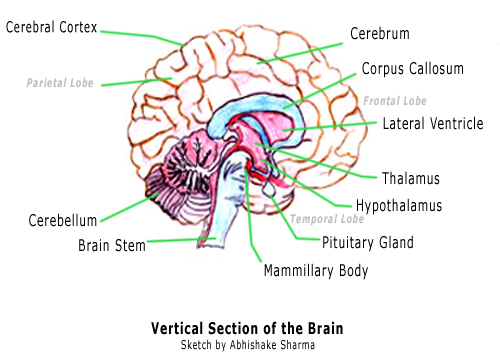
Cerebrum: Anatomy, Function, and Treatment - Verywell Health Corpus callosum: A band of white matter that joins the halves of the cerebrum at the deep center of the brain and coordinates nerve signals between each half. Cerebral arteries: Blood vessels that supply the cerebrum with oxygen-rich blood from the heart. There are three cerebral arteries: anterior (front), middle, and posterior (back).; Circle of Willis: A loop of cerebral arteries and other ...
Stress Effects on the Body: Nervous System
› photos › diagram-of-bodyDiagram Of Body Organs Female Pics Stock Photos, Pictures ... Human internal organs Internal organs in woman and man body. Brain, stomach, heart, kidney, medical icon in female and male silhouette. Digestive, respiratory, cardiovascular systems. Anatomy poster vector illustration. diagram of body organs female pics stock illustrations
Unlabeled Brain Diagram - Cliparts.co
Free Printable Brain Hemisphere Hat - Homeschool Giveaways The brain is one of the most complex and interesting organs in the human body. It is made up of billions of neurons that send messages so that your body can complete different tasks. It communicates through our spinal cord to the different parts of the body. The brain is the most important organ in our body and we be able to live if it wasn't ...

The Most Wonderful Creature: Human Body Organs
Brain Diagram Pons Images, Stock Photos & Vectors | Shutterstock 338 brain diagram pons stock photos, vectors, and illustrations are available royalty-free. See brain diagram pons stock video clips of 4 hypothalamus vector brain diagram with labels labeled brain anatomy pons human body anatomy with labels ventricles in the brain the hypothalamus midbrain cerebellum thalamus Next of 4

Brain anatomy, illustration - Stock Image - C049/3935 - Science Photo Library
Parts of the Brain Activity for Kids, Brain Diagram, and Worksheets for ... Parts of the Brain Worksheet - Label the human brain by writing the number on the brain template Label the Brain Parts Worksheet - Use the brain vocabulary from the word bank to label the brain areas Brain Activity for kids Next we made a 3d brain model for kids using playdough and this brain mold

TooSogiE Medical Images: Cranial Nerves : I - V
Brain: Atlas of human anatomy with MRI - e-Anatomy - IMAIOS MRI Atlas of the Brain. This page presents a comprehensive series of labeled axial, sagittal and coronal images from a normal human brain magnetic resonance imaging exam. This MRI brain cross-sectional anatomy tool serves as a reference atlas to guide radiologists and researchers in the accurate identification of the brain structures.

Found on Bing from www.pinterest.com | Brain anatomy and function, Brain diagram, Brain anatomy
Lateral view of the brain: Anatomy and functions | Kenhub The lateral view of the brain shows the three major parts of the brain: cerebrum, cerebellum and brainstem . A lateral view of the cerebrum is the best perspective to appreciate the lobes of the hemispheres. Each hemisphere is conventionally divided into six lobes, but only four of them are visible from this lateral perspective.

Corticospinal tract - Wikiwand
Left Brain vs. Right Brain: Characteristics Chart [INFOGRAPHIC] Brain dominance theory is absorbing and enjoyable, plus it allows people to think about stereotypes and labels. In reality, though, psychology is complicated, and the truth is that there are very few people who have the traits of only one of these descriptions. More often, we have an intricate combination of both.
Post a Comment for "41 brain diagram and labels"