43 diagram of a cell with labels
Interactive Bacteria Cell Model - CELLS alive In the space are enzymes and other proteins that help digest and move nutrients into the cell. Cell Wall: Composed of peptidoglycan (polysaccharides + protein), the cell wall maintains the overall shape of a bacterial cell. The three primary shapes in bacteria are coccus (spherical), bacillus (rod-shaped) and spirillum (spiral). Structure of Bacterial Cell (With Diagram) - Biology Discussion Cell wall: It is a tough and rigid structure of peptidoglycan with accessory specific materials (e.g. LPS, teichoic acid etc.) surrounding the bacterium like a shell and lies external to the cytoplasmic membrane. It is 10-25 nm in thickness. It gives shape to the cell. Nucleus: The single circular double-stranded chromosome is the bacterial genome.
A Labelled Diagram Of Neuron with Detailed Explanations Diagram Of Neuron. A neuron is a specialized cell, primarily involved in transmitting information through electrical and chemical signals. They are found in the brain, spinal cord and the peripheral nerves. A neuron is also known as the nerve cell. The structure of a neuron varies with their shape and size and it mainly depends upon their ...

Diagram of a cell with labels
Interactive Cell Cycle - CELLS alive INTERPHASE. Gap 0. Gap 1. S Phase. Gap 2. MITOSIS . ^ Cell Cycle Overview Cell Cycle Mitosis > Meiosis > Get the Cell Division PowerPoints Cell Membrane Diagram Labeled : Functions and Diagram Cell Membrane Diagram Labeled Monday, March 22nd 2021. | Diagram Cell Membrane Diagram. There are no organelles in the prokaryotic cells, i.e., they have no internal membrane systems. While lipids help to give membranes their flexibility, proteins monitor and maintain. 03 Label the Cell Diagram | Quizlet Start studying 03 Label the Cell. Learn vocabulary, terms, and more with flashcards, games, and other study tools.
Diagram of a cell with labels. Draw a Neat and Labeled Diagram of a Dry Cell Explanation of the labeling of Dry Cell. The cell is topped with a metal cap which protects the cell. Next, the cell includes a zinc container which is an anode whose base is a negative anode. The dry cell is lined with Porous paper which is a kind of insulating paper, it is a form of Cathode with a carbon rod. The area in between the cathode ... Cells Diagram | Science Illustration Solutions - Edrawsoft Cells Diagram. Cells are the basic building blocks of all living things. The human body is composed of trillions of cells. Cells have many parts, each with a different function. Some of these parts, called organelles, are specialized structures that perform certain tasks within the cell. Drawing cells diagram helps you better understand your ... Structure of Cell: Definition, Types, Diagram, Functions - Embibe Structure of Cell: Cell is the basic functional unit that makes up all living organisms.All organisms, including ourselves, start life as a single cell called the egg. Cells are small microscopic units that perform all essential functions of life and are capable of independent existence. How to Use Cell Values for Excel Chart Labels Select the chart, choose the "Chart Elements" option, click the "Data Labels" arrow, and then "More Options.". Uncheck the "Value" box and check the "Value From Cells" box. Select cells C2:C6 to use for the data label range and then click the "OK" button. The values from these cells are now used for the chart data labels.
Drawing & Labeling a Diagram of a Electrochemical Cell - Study.com Lesson Summary. An electrochemical cell consists of two half-cells connected by a salt bridge.Each half-cell consist of a metal strip in a solution of that metal. Oxidation occurs in one half-cell ... How to draw a nerve cell - labeled science diagrams - YouTube Download a free printable outline of this video and draw along with us: you for watching. Please su... Plant Cell: Diagram, Types and Functions - Embibe Exams Xylem is a tissue that is formed of four different types of cells, i.e. tracheids, xylem vessels, xylem fibres and xylem parenchyma. They are the transport cells in vascular plants. They help in the transport of water and minerals from the roots to the leaves and other parts of the plants. The movement of water is unidirectional. Phloem Galvanic Cell: Definition, Diagram and Working - Science ABC Jan 17, 2022 · Galvanic Cell vs Electrolytic Cell. Lastly, once dead, galvanic cells cannot be revived or recharged. This is why one must change the batteries in an alarm clock or remote control from time to time. The kind of electrochemical cell that can be recharged is an electrolytic cell.
How to plot a ternary diagram in Excel - Chemostratigraphy.com Feb 13, 2022 · Adding labels to the apices. Next, we need some space for the apices labels: click into the Plot Area (not the Chart Area) then resize by holding the Shift key (this ensures an equal scaling) and use the mouse cursor on one of the corner pick-points. Then recentre the Plot Area in the Chart Area. Animal Cell Diagram with Label and Explanation: Cell Structure, Functions Diagram of Animal Cell Below is the diagram of the animal cell which shows the organelles present in it. The cell is covered with cytoplasm which consists of cell organelles in it. The nucleus is covered with a rough Endoplasmic Reticulum and other organelles each designed for a specific purpose. Cell: Structure and Functions (With Diagram) - Biology Discussion Eukaryotic Cells: 1. Eukaryotes are sophisticated cells with a well defined nucleus and cell organelles. 2. The cells are comparatively larger in size (10-100 μm). 3. Unicellular to multicellular in nature and evolved ~1 billion years ago. 4. The cell membrane is semipermeable and flexible. 5. These cells reproduce both asexually and sexually. Plant Cell Diagram | Science Trends A plant cell diagram, like the one above, shows each part of the plant cell including the chloroplast, cell wall, plasma membrane, nucleus, mitochondria, ribosomes, etc. A plant cell diagram is a great way to learn the different components of the cell for your upcoming exam. Plants are able to do something animals can't: photosynthesize.
Bipolaris sp.
Learn the parts of a cell with diagrams and cell quizzes For this exercise we'll start with an image of a cell diagram ready labeled. Study this and make sure that you're clear about which structure is found where. Cell diagram unlabeled It's time to label the cell yourself! As you fill in the cell structure worksheet, remember the functions of each part of the cell that you learned in the video.
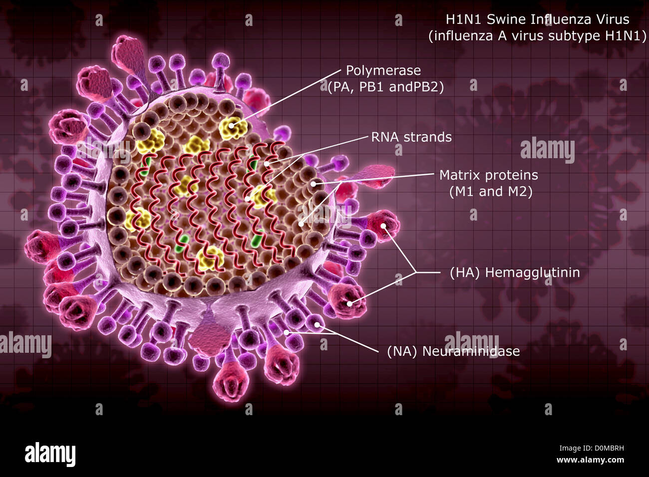
Diagram of an H1N1 swine flu virus particle with section labels Stock Photo - Alamy
Animal Cells: Labelled Diagram, Definitions, and Structure Animal Cells Organelles and Functions. A double layer that supports and protects the cell. Allows materials in and out. The control center of the cell. Nucleus contains majority of cell's the DNA. Popularly known as the "Powerhouse". Breaks down food to produce energy in the form of ATP.
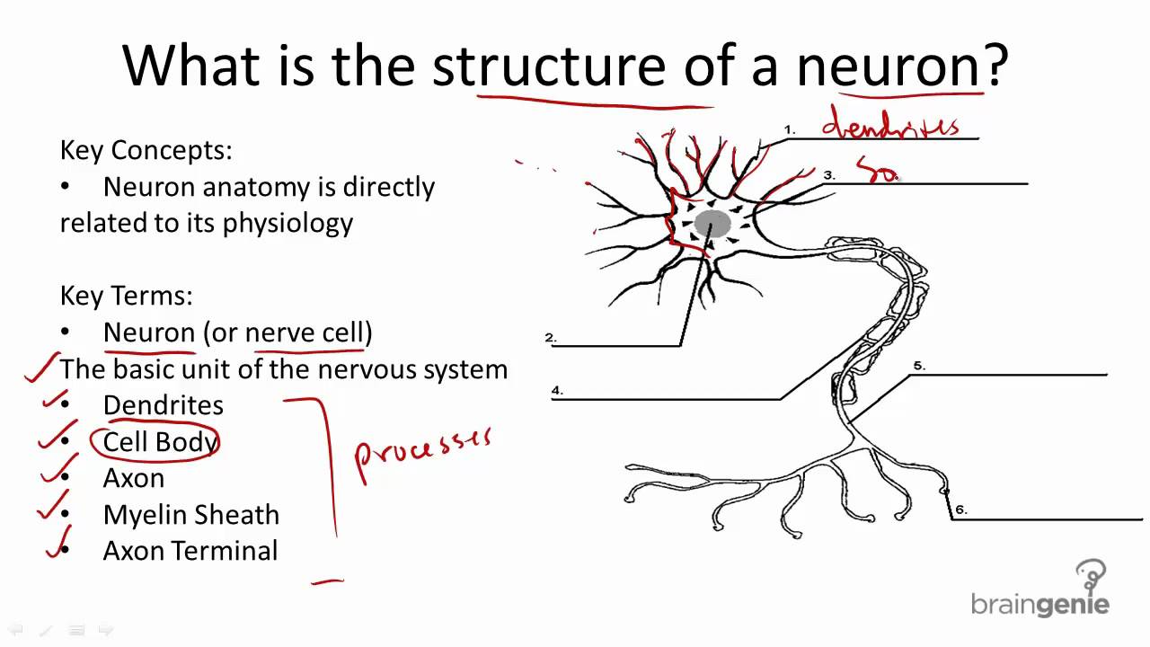
8 1 3 Nerve Cell Structure and Function - YouTube
Label the cell - Teaching resources Label Plant and Animal Cell Labelled diagram by Sciencegeek 5.6 Label the sentence Labelled diagram by Christianjolene the cell Match up by Elenagp9149 5.7 Label the sentence Labelled diagram by Christianjolene The Cell Labelled diagram by U27704813 Label the Electromagnetic Spectrum Labelled diagram by Elizabetheck G6 G7 G8 Science

Pinus Stem Cross Section Labeled - Top Label Maker
Human Cell Diagram, Parts, Pictures, Structure and Functions Diagram of the human cell illustrating the different parts of the cell. Cell Membrane. The cell membrane is the outer coating of the cell and contains the cytoplasm, substances within it and the organelle. It is a double-layered membrane composed of proteins and lipids. The lipid molecules on the outer and inner part (lipid bilayer) allow it to ...
Post a Comment for "43 diagram of a cell with labels"